Home
/ Anatomy Of Your Back Organs - Thoracic Spine Anatomy And Upper Back Pain : Although the majority of back pain episodes are caused by injury to one or more of the structures of the lumbar spine, there are several conditions affecting the internal organs that may also result in symptoms of back pain.
Anatomy Of Your Back Organs - Thoracic Spine Anatomy And Upper Back Pain : Although the majority of back pain episodes are caused by injury to one or more of the structures of the lumbar spine, there are several conditions affecting the internal organs that may also result in symptoms of back pain.
Anatomy Of Your Back Organs - Thoracic Spine Anatomy And Upper Back Pain : Although the majority of back pain episodes are caused by injury to one or more of the structures of the lumbar spine, there are several conditions affecting the internal organs that may also result in symptoms of back pain.. Although the majority of back pain episodes are caused by injury to one or more of the structures of the lumbar spine, there are several conditions affecting the internal organs that may also result in symptoms of back pain. 7 photos of the human body organs in lower back. The back supports the weight of the body, allowing for flexible movement while protecting vital organs and nerve structures. The anatomy of the back refers to the muscles of the back, as well as the bones of the scapulae, ribcage, and spine. Human body anatomy female female anatomy muscle shoulder blade pain anatomy back muscles bones man female anatomy body muscles in a body female anatomy muscole shoulder concept muscular sysyem.
Each are symmetrically paired on a right and left side. The normal arrangement of internal organs is known as situs solitus.although cardiac problems are more common, many people with situs inversus have no medical symptoms or complications resulting from the condition, and. The back muscles enable you to stand up straight; The lower spine, the hips and tailbone, and the abdomen. The left lumbar region is one of nine regions of the abdominal cavity, and it contains organs from both the digestive and excretory systems.
Internal Organs Back View Stock Illustrations 86 Internal Organs Back View Stock Illustrations Vectors Clipart Dreamstime from thumbs.dreamstime.com The muscles of the lower back, including the erector spinae and quadratus lumborum muscles, contract to extend and laterally bend the vertebral column. The outer portion contains neurons, and the inner area communicates with the cerebral cortex. The back anatomy includes some of the most massive and functionally important muscles in the human body. The vertebral column runs the length of the back and creates a central area of recession. Support and protect your spine; 7 photos of the human body organs in lower back. The nerves in your thoracic spine go to your chest and abdomen. The back supports the weight of the body, allowing for flexible movement while protecting vital organs and nerve structures.
And reach, pull and extend your arms and torso.
Lumbar spine anatomy video understanding the anatomy of your lower spine can help you communicate more effectively with the medical professionals who treat your lower back pain. And reach, pull and extend your arms and torso. The muscles of the lower back, including the erector spinae and quadratus lumborum muscles, contract to extend and laterally bend the vertebral column. Five vital organs that are essential for survival are the brain, heart, kidneys, liver, and lungs. Still, many individuals pay far too little attention to them. In the bladder diagram above, you can see the structure of the urinary bladder. The back functions are many, such as to house and protect the spinal cord, hold the body and head upright, and adjust the movements of the upper and lower limbs. Just below the diaphragm is the liver and the gall bladder. Although the majority of back pain episodes are caused by injury to one or more of the structures of the lumbar spine, there are several conditions affecting the internal organs that may also result in symptoms of back pain. The nerves of the lumbar spine then reach to your legs, bowel, and bladder. The normal arrangement of internal organs is known as situs solitus.although cardiac problems are more common, many people with situs inversus have no medical symptoms or complications resulting from the condition, and. Your back consists of a complex array of bones, discs, nerves, joints, and muscles. The human rib cage is made up of 12 paired rib bones;
The major organs of the abdomen include the. 3d illustration showing organs of digestive system with highlighted pancreatic gland, view from back ribs with nerves posterior view. Female cardiovascular system, rear and front views, on black. The human rib cage is made up of 12 paired rib bones; And reach, pull and extend your arms and torso.
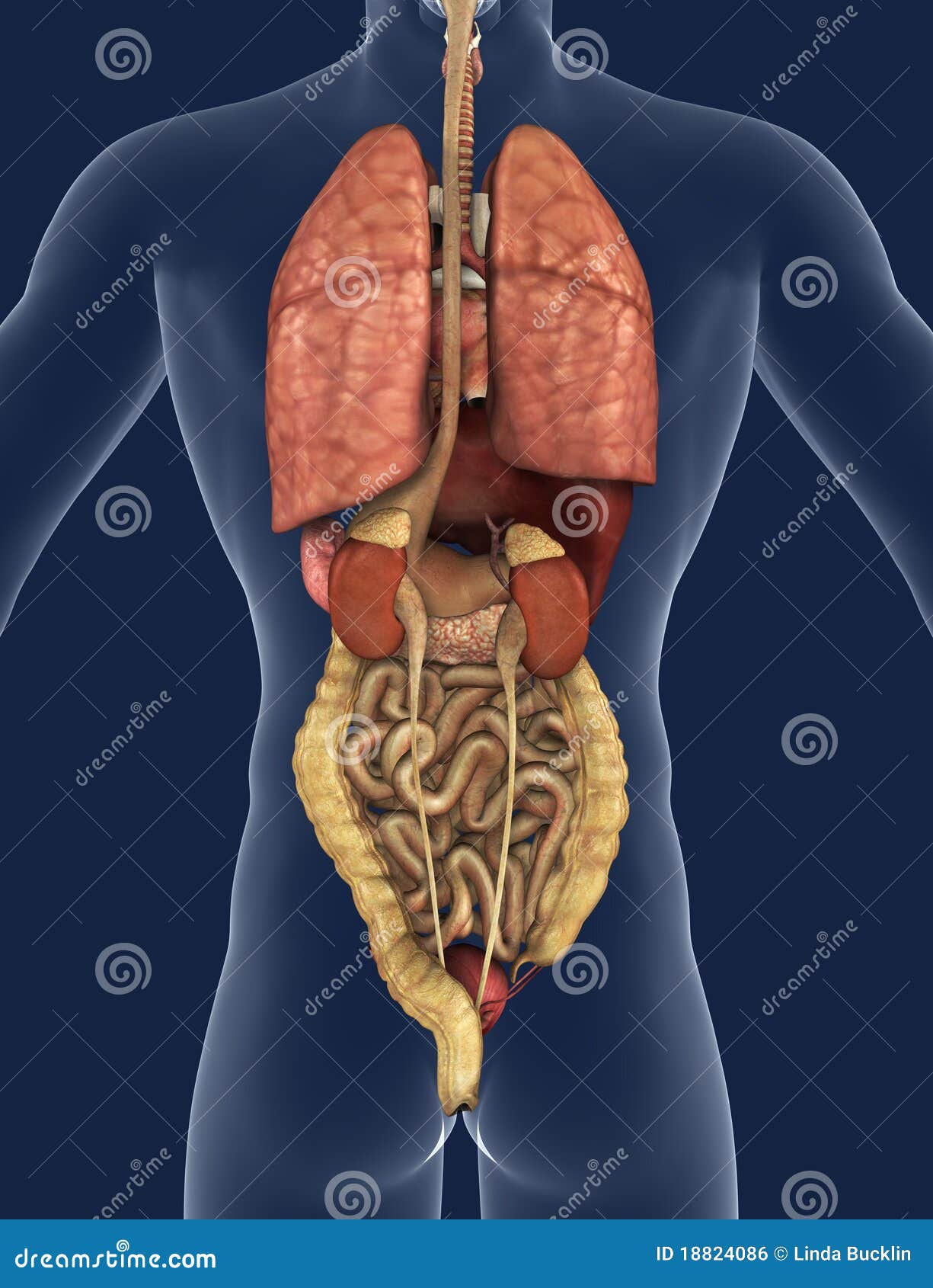
Internal Organs Back View Stock Illustration Illustration Of Posterior 18824086 from thumbs.dreamstime.com The abdominal muscles also play a major role in the posture and stability to the body and compress the organs of the abdominal cavity during various activities such as breathing and defecation. Situs inversus (also called situs transversus or oppositus) is a congenital condition in which the major visceral organs are reversed or mirrored from their normal positions. The lumbar region of the spine, more commonly known as the lower back, is situated between the thoracic, or chest, region of the spine, and the sacrum. Lumbar spine anatomy video understanding the anatomy of your lower spine can help you communicate more effectively with the medical professionals who treat your lower back pain. Support and protect your spine; The major muscles of the abdomen include the rectus abdominis in front, the external obliques at the sides, and the latissimus dorsi muscles in the back. Covering an expanse from the neck to the tailbone, the back muscles are responsible for a broad range of functions, from extending the spine to shrugging the shoulders. The back is the body region between the neck and the gluteal regions.
The back is the body region between the neck and the gluteal regions.
If you want to keep your back healthy & functional, you'll want to sit less, move more, & do these 7 lower back stretches. In my 25 years plus of teaching experience i have found that exercise, pilates, yoga, and movement will really help some clients with back pain. See human back anatomy stock video clips. The lower back is really composed of three areas of the body: The back muscles enable you to stand up straight; The major organs of the abdomen include the. The muscles of your back support your spine, attach your pelvis and shoulders to your trunk, and provide mobility and stability to your trunk and spine. Support and protect your spine; Since the spine is encircled by musculature, the abdomen, spinal muscles, and hips are all integral aspect of maintaining a healthy lower spine and therefore lower back. The nerves in your thoracic spine go to your chest and abdomen. Browse 384 human anatomy organs back view stock photos and images available, or start a new search to explore more stock photos and images. At the level of the pelvic bones, the abdomen ends and the pelvis begins. The human rib cage is made up of 12 paired rib bones;
It is the surface of the body opposite from the chest and the abdomen. If you want to keep your back healthy & functional, you'll want to sit less, move more, & do these 7 lower back stretches. Spleen is 1 inch thick, 3 inches broad and 5 inches long. The lumbar region of the spine, more commonly known as the lower back, is situated between the thoracic, or chest, region of the spine, and the sacrum. Human anatomy · july 23, 2016.
Internal Organs Back View Stock Illustrations 86 Internal Organs Back View Stock Illustrations Vectors Clipart Dreamstime from thumbs.dreamstime.com The outer portion contains neurons, and the inner area communicates with the cerebral cortex. The human rib cage is made up of 12 paired rib bones; Still, many individuals pay far too little attention to them. The back anatomy includes some of the most massive and functionally important muscles in the human body. Human anatomy female human anatomy picture human anatomy chart human anatomy and physiology body anatomy organs human body organs human body parts human organ diagram. The brain is one of the largest and most complex organs in the human body. Although the majority of back pain episodes are caused by injury to one or more of the structures of the lumbar spine, there are several conditions affecting the internal organs that may also result in symptoms of back pain. If you want to keep your back healthy & functional, you'll want to sit less, move more, & do these 7 lower back stretches.
Browse 384 human anatomy organs back view stock photos and images available, or start a new search to explore more stock photos and images.
Just below the diaphragm is the liver and the gall bladder. The lumbar region of the spine, more commonly known as the lower back, is situated between the thoracic, or chest, region of the spine, and the sacrum. Each are symmetrically paired on a right and left side. The outer portion contains neurons, and the inner area communicates with the cerebral cortex. The vertebral column runs the length of the back and creates a central area of recession. The human back, also called the dorsum, is the large posterior area of the human body, rising from the top of the buttocks to the back of the neck. Still, many individuals pay far too little attention to them. Five vital organs that are essential for survival are the brain, heart, kidneys, liver, and lungs. In the bladder diagram above, you can see the structure of the urinary bladder. Spleen is 1 inch thick, 3 inches broad and 5 inches long. 3d illustration showing organs of digestive system with highlighted pancreatic gland, view from back ribs with nerves posterior view. The nerves also carry electrical signals back to the brain that allow you to feel sensations. The human rib cage is made up of 12 paired rib bones;